Anatomical reminder
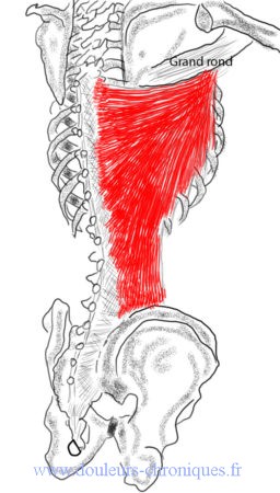
This very extensive muscle is inserted, in its medial part, on the spines of the last 6 thoracic vertebrae, the 5 lumbar vertebrae, on the sacrum, on the iliac crest and on the last ribs (3 or 4). In its lateral part, it fits on the anterior surface of the proximal part of the humerus with the teres major.
Its function is adduction and internal rotation of the arm.
Latissimus dorsi muscle myofascial syndrome
Description
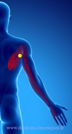
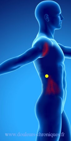
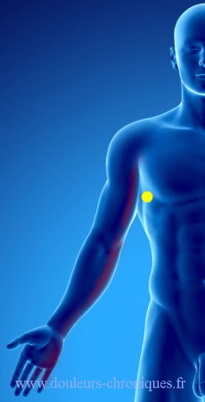
In yellow, the trigger point
In red, referred pain
The referred pains are mainly located in the posterolateral chest wall at the tip of the scapula, they radiate into the arm on its posterior and internal surfaces up to the 4th and 5th fingers. Pain from the side of the abdomen to the iliac crest is also possible
Triggering factor
The repeated upward and forward movements of the arm, especially when lifting weights, or even pulling objects downwards.
Diagnostic
Palpation of the painful points by “pinching” the muscle next to the intermediate part of the scapula reproduces the pain. Myofascial syndrome of the latissimus dorsi muscle leads to limited movement of the shoulder
Treatment
- Patient lying on back, stretching arm up and slightly sideways
- patient lying in contralateral lateral decubitus, arm up elbow flexed in complete abduction
- Infiltration of lidocaine in the part of the mid-thoracic muscle followed by stretching (described above)
Self-stretching by “covering the mouth” exercise and stretching exercise in “a doorway” - Self-stretching by “covering the mouth” exercise and stretching exercise in “a doorway”